MEDICAL / SURGICAL RETINA & LASERS

This Includes Myriads of Retinal disorder including Diabetic Retinopathy, Retinal detachment,Age related maculopathy (AMD), Macular holes, Artery and Vein Occlusions and many.
TREATMENT:
Laser therapy and Surgical Correction
What is Diabetic Retinopathy?
 Diabetic Retinopathy is an eye disease affecting the retina (the black portion of the eye) and is a frequent complication of diabetic disease. Diabetes damages the small blood vessels in the retina of the eye and can lead to poor vision and even blindness over a period of time.
During the early stages, the tiny blood vessels in the eye weaken and develop small bulges that may burst (causing haemorrhage) and leak into the retina (causing muscular edema) and also into the gel-like fluid inside the eye called the vitreous gel (causing vitreous haemorrhage). This causes visual deterioration. As the condition progresses, new fragile blood vessels grow on the surface of the retina, impairing vision. All these changes can cause permanent visual deterioration and loss. This is called Diabetic Retinopathy.
There is no retina surgery & treatment that can prevent diabetic retinopathy altogether. People usually with any form of diabetes may develop diabetic retinopathy. However, with regular exercising and keeping your sugar levels under control it has been proven that diabetic retinopathy’s progress slows down considerably.
Over the years a variety of retina treatments have sprung up to cure this disease such as scatter photocoagulation, focal photocoagulation, and vitrectomy which prevent blindness in most people. The sooner retinopathy is diagnosed, the more likely these treatments will be successful. And that is why you need the expert help of DOVS , a pioneering name in the Diabetic Retinopathy in Mumbai, India.
Retinal Detachment
Retinal detachment is a separation of the light-sensitive membrane in the back of the eye (the retina) from its supporting layers.
Causes, incidence, and risk factors
The retina is the clear tissue in the back of the eye. It helps you see the images that are focused on it by the cornea and the lens.
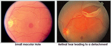
- The most common type of retinal detachments is often due to a tear or hole in the retina. Eye fluids may leak through this opening. This causes the retina to separate from the underlying tissues, much like a bubble under wallpaper. This is most often caused by a condition called posterior vitreous detachment. However, it may also be caused by trauma and very bad nearsightedness (high minus power). A family history of retinal detachment also increases your risk.
- Another type of retinal detachment is called tractional detachment. This is seen in people who have uncontrolled diabetes, previous retinal surgery, or have chronic inflammation.
When the retina becomes detached, bleeding from area blood vessels may cloud the inside of the eye, which is normally filled with vitreous fluid. Central vision becomes severely affected if the macula, the part of the retina responsible for fine vision, becomes detached.
Symptoms
Retinal detachment is generally preceded by the formation of holes(s) or tears in the retina; symptoms of which may include sudden onset of flashes and floaters-multiple black spots or cobweb like floating objects in front of the affected eye.
- When detachment occurs, appearance of curtain falling in front of the eye, decreased vision or total obscuration of vision.
- Bright flashes of light, especially in peripheral vision
- Blurred vision
- Floaters in the eye
- Sudden appearance of Shadow or blindness in a part of the visual field of one eye
Diabetic Eye Diseases
 Diabetes and eye diseases go hand in hand. As a matter of fact, one of the major concerns for diabetic patients is that they might lose their eyesight if their sugar levels are not kept under control for long. A question raging in the minds of people might be how can diabetes which is a disease of the pancreas can affect such a dire affect over your vision?
This can be simply explained with the occurrence of diabetes in our blood stream. When we eat food, it gives us glucose which when broken by the natural insulin of our body releases small cells of energy. In a diabetic patient, his natural insulin mechanism fails and the unbroken glucose gets absorbed in our blood stream thus causing elevated levels of sugar in our body.
You may have heard that diabetes causes eye problems and may lead to blindness. People with diabetes do have a higher risk of permanent blindness and other various eye problems than people without diabetes. Diabetes can affect the eyes and vision in a number of ways. It may lead to frequent fluctuations in vision, cataract in young age, decreased vision due to involvement of optic nerve, temporary paralysis of the muscles controlling the movement of eyes and thus double vision, the Incidence glaucoma is higher in diabetics. The most significant complication of diabetes in eye is diabetic retinopathy. This may cause irreversible loss of vision in later stages.
Age Related Macular Degeneration
Get affordable world class Macular Degeneration Treatment in @ DOVS!
Age-related Macular Degeneration (AMD or ARMD) is a medical condition which usually affects older adults and results in a loss of vision in the center of the visual field (the macula) because of the damage done to the retina in this region. It occurs in both “dry” and “wet” forms and is considered to be a major cause of blindness and visual impairment in older adults (>50 years). Macular degeneration can make it difficult or impossible to read or recognize faces, although some peripheral vision remains to allow other activities of daily life.
The loss of central vision profoundly affects visual functioning. It is quite difficult, for example, to read without central vision. Pictures that attempt to depict the central visual loss of macular degeneration with a black spot do not really do justice to the devastating nature of the visual loss. This can be demonstrated by printing letters six inches high on a piece of paper and attempting to identify them while looking straight ahead and holding the paper slightly to the side. Most people find this quite difficult to perform..
There is a loss of contrast sensitivity, so that contours, shadows, and color vision are less vivid. The loss in contrast sensitivity can be quickly and easily measured by a contrast sensitivity test performed by an eye specialist.
This loss of central vision makes day to day working quite difficult for the affected individual.
For more updates about Eye Care, visit Department of Visual Sciences and Eye Donation Center Facebook Page