CORNEA AND OCULAR SURFACE
Cornea is the hour glass structure at the front of the eye. . It provides about 2/3 of the eye’s focusing power. It gives us a clear window to look through and is the key to good vision. . It also helps shield the rest of the eye from germs, dust, and other harmful pollutants.
Infections, chemical burns and injuries cause this crystal clear structure to opacify. Also conditions like Keratoconus and dystrophies affect the focusing ability of the cornea.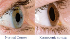
Treatments:
Replacement of the cornea with a freshly donated cornea from the cadaver.this procedure is called Keratoplasty.
Use of artificial cornea is called Keratoprosthesis and may be used in multiple graft failures.The Ocular Surface can be reconstructed using aminiotic membrance and stem cells.
The Corneal Service at our clinic provides medical and surgical care to a wide variety of corneal, external eye related diseases and anterior segment eye disorders and corneal refractive surgery.
Cornea & External eye diseases:
- Corneal Infections
- Ocular surface trauma
- Ocular surface disorders like blepharitis, meibomitis, conjunctival and scleral diseases
- Refractive errors (nearsightedness, farsightedness and astigmatism)
- Conjunctivitis (red eye)
- Dry eye syndrome
- Ectatic corneal diseases like keratoconus, keratoglobus and pellucid marginal degeneration
- Pterygium
- Corneal dystrophies and degenerations
- Eye allergies Corneal complications arising from other forms of ocular surgery
What is Keratoprosthesis?
Cornea/Keratoconus
In patients with keratoconus the cornea becomes cone shaped (hence the name keratoconus, derived from the Greek word for cornea (‘kerato’) and cone shaped (‘conus’). In patients with keratoconus the cornea is not only cone shaped but the surface is also irregular resulting in a distorted image being projected onto the brain, this is usually a progressive condition which at times can lead to permanent loss of vision if scaring occurs, but in most cases can cause partial loss or visual distortion.
In extreme cases there often is no remedy but a complete transplant of the cornea with a lengthy surgical procedure.
Keratoconus is often discovered during adolescence. In late stage it can usually be diagnosed with slit-lamp examination of the cornea. But in initial stages it can be very easily missed and can be diagnosed with more accurate test is called corneal topography, which creates a map of the curve of the cornea.
When keratoconus is advanced, the cornea will be thinner at the point of the cone. This can be measured with a painless test called pachymetry. By methods such as Orbscan / Pentacam or Sirius machines.
Because the cornea is irregular and cone shaped, glasses do not adequately correct the vision in patients with keratoconus since they cannot conform to the shape of the eye. Patients with keratoconus see best with rigid contact lenses since these lenses provide a clear surface in front of the cornea allowing the light rays to be projected clearly to the retina. These patients are treated with rigid contact lenses. , But these rigid contact lenses may not be well tolerated by most of the patients.
There is however some excellent new surgical options for patients with keratoconus who cannot tolerate these lenses, these options are discussed under treatments for keratoconus, Corneal Cross linking or CXL or even C3R. Many patients are initially unaware they have keratoconus and see their eye doctor because of increasing spectacle blur or progressive changes in their prescription. Classically my present in change in cylindrical power or axis in the prescription glasses.
Keratoconus typically commences at puberty and progresses to the mid thirties at which time progression slows and often stops. Between age 12 and 35 it can arrest or progress at any time and there is now way to predict how fast it will progress or if it will progress at all. In general young patients with advanced disease are more likely to progress to the point where they may ultimately require some form of surgical intervention.
What is Keratoplasty
Keratoconus is a disease that causes a progressive thinning of the cornea (the clear front portion of the eye). As a result of this condition, the normal outward pressure from within the eye causes the cornea to progressively bulge into a cone-like shape.
What is Ocular Surface?
Treatment of these conditions involve ocular surface reconstruction. This involves multidisciplinary approach to restore the normal ocular surface defense so that a stable tear film can be achieved, restore the epithelial stem cells and restore stem cell stromal niche. These treatment strategies combine medical and surgical modalities. Medical treatment includes the use of various topical drops. Surgical modalities include symblepharon release, pannus excision, amniotic membrane transplantation, conjunctival autograft, limbal stem cell transplant etc.
For more updates about Eye Care, visit Department of Visual Sciences and Eye Donation Center Facebook Page